A Prospective Study to Find the Relationship of Carotid Stenosis and Plaque Morphology with Infarct Volume in Ischemic Stroke Patients
Authors
##plugins.themes.bootstrap3.article.main##
Abstract
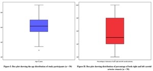
Background: Ischemic stroke, a major global health concern, is often caused by carotid artery stenosis resulting from atherosclerotic plaque formation. Carotid Doppler ultrasonography is a non-invasive method to assess the degree of stenosis and the characteristics of plaques, which are crucial in evaluating stroke risk and guiding treatment decisions. Objective: This study aimed to explore the relationship between the percentage of carotid artery stenosis, plaque morphology, and infarct volume in patients with acute ischemic stroke. Design: Cross-sectional analysis. Subjects/Patients: Fifty patients diagnosed with ischemic stroke at the Department of Radiodiagnosis, PSG Institute of Medical Sciences and Research, Coimbatore, between January 2019 and June 2020. Methods: Carotid Doppler ultrasound was used to quantify stenosis and classify plaques based on echogenicity, surface contour, and internal texture. Infarct volumes were measured using MRI. Statistical correlations and regression analyses were conducted to identify significant relationships. Results: Carotid stenosis was observed bilaterally in half of the cohort, with average stenosis of 62.2% (right) and 55.4% (left). A moderate positive correlation was found between stenosis percentage and infarct volume (r=0.446, p=0.035). Plaque morphology did not significantly correlate with infarct size. Regression analysis showed that age and stenosis severity accounted for 48% of infarct volume variation. Conclusion: Carotid stenosis severity is a more reliable predictor of infarct volume than plaque morphology, highlighting the value of Doppler-based stenosis assessment in ischemic stroke management.
##plugins.themes.bootstrap3.article.details##
Copyright (c) 2025 S Thilagarajan, MD, Sumana Kedilaya, MD, Senthil Prabu M

This work is licensed under a Creative Commons Attribution 4.0 International License.
Creative Commons License All articles published in Annals of Medicine and Medical Sciences are licensed under a Creative Commons Attribution 4.0 International License.
[1] Truelsen T, Begg S, Mathers C. The global burden of cerebrovascular disease. Global burden of disease 2000. Cerebrovascular disease. 2006.
[2] Ojaghihaghighi S, Vahdati SS, Mikaeilpour A, Ramouz A. Comparison of neurological manifestation in patients with hemorrhagic and ischemic stroke. World J Emerg Med 2017; 8: 34-38.
[3] Sztajzel R. Ultrasonographic assessment of the morphological characteristics of the carotid plaque. Swiss Med Wkly 2005; 135: 635-643.
[4] Demarco JK, Ota H, Underhill HR, Zhu DC, Reeves MJ, Potchen MJ, et al. MR carotid plaque imaging and contrast-enhanced MR angiography identifies lesions associated with recent ipsilateral thromboembolic symptoms: An in vivo study at 3 T. Am J Neuroradiol 2010; 31: 1395-1402.
[5] Alagoz AN, Acar BA, Acar T, Karacan A, Demiryurek BE. Relationship between carotid stenosis and infarct volume in ischemic stroke patients. Med Sci Monit 2016; 22: 4954-4959.
[6] Zhao H, Zhao X, Liu X, Cao Y, Hippe DS, Sun J, et al. Association of carotid atherosclerotic plaque features with acute ischemic stroke: A magnetic resonance imaging study. Eur J Radiol 2013; 82: e564-e570.
[7] Ahmed L, Noori FA. Carotid Doppler study in patients with cerebral infarction. Fac Med Baghdad 2011; 53: 47-51.
[8] Harrigan MR, Deveikis JP. Contemporary Medical Imaging. In: Handbook of Cerebrovascular Disease and Neurointerventional Technique. New York: Springer; 2009. p. 3.
[9] Shapiro M, Becske T, Riina HA, Raz E, Zumofen D, Jafar JJ, et al. Toward an Endovascular Internal Carotid Artery Classification System. Am J Neuroradiol 2014; 35: 230-236.
[10] Wolpert SM. The circle of Willis. Am J Neuroradiol 1997; 18: 1033-1034.
[11] Osborn AG. Diagnostic Cerebral Angiography. 2nd ed. Philadelphia: Lippincott; 1999.
[12] Alpers BJ, Berry RG. Circle of Willis in cerebral vascular disorders: The anatomical structure. Arch Neurol 1963; 8: 398-402.
[13] Marinkovic S, Milisavljevic M, Marinkovic Z. Branches of the anterior communicating artery. Microsurgical anatomy. Acta Neurochir (Wien) 1990; 106: 78-85.
[14] Gloger S, Gloger A, Vogt H, Kretschmann HJ. Computer-assisted 3D reconstruction of the terminal branches of the cerebral arteries. I. Anterior cerebral artery. Neuroradiology 1994; 36: 173-180.
[15] Harrigan MR, Deveikis JP. Contemporary Medical Imaging. In: Handbook of Cerebrovascular Disease and Neurointerventional Technique. New York: Springer; 2009. p. 49.
[16] Gibo H, Lenkey C, Rhoton AL Jr. Microsurgical anatomy of the supraclinoid portion of the internal carotid artery. J Neurosurg 1981; 55: 560-574.
[17] Djulejic V, Marinkovic S, Georgievski B, Stijak L, Aksic M, Puskas L, et al. Clinical significance of blood supply to the internal capsule and basal ganglia. J Clin Neurosci 2016; 25: 19-26.
[18] Duan S, He H, Shaomao LV, Chen L. Three-dimensional CT study on the anatomy of vertebral artery at atlantoaxial and intracranial segment. Surg Radiol Anat 2010; 32: 39-44.
[19] Balyasnikova S, Fischer BM, Nijs RB. PET/MR in oncology: An introduction with focus on MR and future perspectives for hybrid imaging. Am J Nucl Med Mol Imaging 2012; 2: 458-474.
[20] Lee W. General principles of carotid Doppler ultrasonography. Ultrasonography 2014; 33: 11-17.
[21] Aljubawii AAAA, Hussein HN, Al-Alwany AA. Prevalence of coronary artery disease in patients with left bundle branch block and its association with risk factors hypertension and diabetes mellitus. Med J Babylon 2023; 20: 867-870.
[22] Alabdaly MA, Ahmed HM, Thanoun BI, Al Ashow SAM, Ahmad WG. Frequency of stroke and distribution of its subtypes in Nineveh, Iraq, related to risk factors. Med J Babylon 2024; 21(Suppl 2): S234-S237.
[23] Aljubawii AAA, Ali AI, Al Mamuri HAE. Relationship between modifiable atherosclerotic cardiovascular risk factors and coronary artery bifurcation lesion. Med J Babylon 2023; 20: 777-783. doi: 10.4103/MJBL.MJBL_481_23.
[24] Natarajan I, Rajendran K, Narayanan S, Shanmugam J. Swachh Bharat Mission Gramin: Uptake and challenges in rural Coimbatore. J Fam Med Prim Care 2024; 13: 4539-4544. doi: 10.4103/jfmpc.jfmpc_91_24.
[25] Charles J, Rajendran K, Osborn J. An Internal Audit on Patient Safety and Quality of services in a Rural Health Training Center, Coimbatore. Digit J Clin Med 2024; 6: [no page]. doi:10.55691/2582-3868.1175.
[26] Balasundararaj D, Prabhu MS, Thilagarajan S, Rajendran K. Analysis of Preanalytical Errors in Clinical Biochemistry Laboratory of a Tertiary Care Hospital: A Retrospective Study. J Clin Diagn Res 2024; 18: BC06-BC09. doi:10.7860/JCDR/2024/73364/20384.

