
Risk Stratification for Thyroid Incidentalomas on PET/CT: Systematic Review and Meta-Analysis
Authors

##plugins.themes.bootstrap3.article.sidebar##
##plugins.themes.bootstrap3.article.main##
Abstract
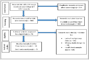
Background: Focal thyroid incidentalomas (FTIs) found on 18F-FDG PET/CT (Fluorine-18-Fluorodeoxyglucose Positoin Emission Tomography/Computed Tomography) examinations are becoming commonplace in routine clinical practice. While in most situations benign, a high percentage.Objective and Aims: Establish the pooled prevalence and risk of malignancy of FTIs on PET/CT and define imaging and clinical risk markers of malignancy. Research questions: What is the malignant rate of FTIs recognized with 18F-FDG PET/CT, and what risk stratification factors are?Methods: PubMed, Scopus, and Web of Science were searched extensively to compile studies between 2015 and 2025. Included were studies that reported sample size and results in relation to malignancy. Data extraction and quality assessment were carried out with the Newcastle-Ottawa Scale. Random-effects meta-analysis was applied to establish pooled prevalence and 95% confidence intervals.Results: Ten studies with 2,500 patients were reviewed. The combined prevalence of FTIs was 2.5%, with an overall risk of malignancy of 30%. Elevated SUVmax (standardized uptake value), suspicious ultrasound morphology, and cytologic proof were reliable predictors of malignancy.Conclusion: FTIs on PET/CT carry high malignancy risk, necessitating systematic evaluation with combined metabolic, morphologic, and cytologic analysis. Reporting and risk stratification guidelines should be established. Prospective research should focus on development of AI-based predictive models and machine learning algorithms to enhance early detection and reduce unnecessary procedures.
##plugins.themes.bootstrap3.article.details##
Copyright (c) 2025 Shamim Hyder, Nadha Rahim, Noula Rahim, Jamila Hameed

This work is licensed under a Creative Commons Attribution 4.0 International License.
Creative Commons License All articles published in Annals of Medicine and Medical Sciences are licensed under a Creative Commons Attribution 4.0 International License.
Shamim Hyder, Department of Radiology, Karuna Medical College, Vilayodi, Chittur, Palakkad, Kerala, India.
Department of Radiology, Karuna Medical College, Vilayodi, Chittur, Palakkad, Kerala, India.
Nadha Rahim, Department of Radiology, Karuna Medical College, Vilayodi, Chittur, Palakkad, Kerala, India.
Department of Radiology, Karuna Medical College, Vilayodi, Chittur, Palakkad, Kerala, India.
Noula Rahim, Department of Biochemistry, Karuna Medical College, Vilayodi, Chittur, Palakkad, Kerala, India.
Department of Biochemistry, Karuna Medical College, Vilayodi, Chittur, Palakkad, Kerala, India.
Jamila Hameed, Research Mentor, Karuna Medical College, Vilayodi, Chittur, Palakkad, Kerala, India.
Research Mentor, Karuna Medical College, Vilayodi, Chittur, Palakkad, Kerala, India.
[1] Cooper DS, Doherty GM, Haugen BR, et al; for the American Thyroid Association Guidelines Taskforce on Thyroid Nodules and Differentiated Thyroid
[2] Cancer. Revised American Thyroid Association management guidelines for
[3] patients with thyroid nodules and differentiated thyroid cancer. Thyroid. 2009;
[4] 19:1167-1214.
[5] Eren MŞ, Yılmaz A, Kılıçkesmez Ö, Yalçın B, Yılmaz M, Kızılkaya M, et al. The incidence of 18F-FDG PET/CT thyroid incidentalomas and the role of SUVmax in the differentiation of benign and malignant lesions. Nucl Med Commun. 2016;37(12):1219–25.
[6] Meyer HJ, Wienke A, Surov A. Associations between GLUT expression and SUV values derived from FDG-PET in different tumors—A systematic review and meta analysis. PloS one. 2019 Jun 17;14(6):e0217781.
[7] de Koster EJ, de Geus-Oei LF, Brouwers AH, van Dam EW, Dijkhorst-Oei LT, van Engen-van Grunsven AC, van den Hout WB, Klooker TK, Netea-Maier RT, Snel M, Oyen WJ. [18F] FDG-PET/CT to prevent futile surgery in indeterminate thyroid nodules: a blinded, randomised controlled multicentre trial. European Journal of Nuclear Medicine and Molecular Imaging. 2022 May;49(6):1970-84.
[8] Jamsek J, Zagar I, Gaberscek S, Grmek M. Thyroid lesions incidentally detected by (18)F-FDG PET-CT - a two centre retrospective study. Radiol Oncol. 2015;49(2):121-127. Published 2015 Mar 25.
[9] Pattison DA, Bozin M, Gorelik A, Hofman MS, Hicks RJ, Skandarajah A. 18F-FDG–avid thyroid incidentalomas: the importance of contextual interpretation. Journal of Nuclear Medicine. 2018 May 1;59(5):749-55.
[10] Agrawal K, Weaver J, Ul-Hassan F, Jeannon JP, Simo R, Carroll P, Hubbard JG, Chandra A, Mohan HK. Incidence and Significance of Incidental Focal Thyroid Uptake on (18)F-FDG PET Study in a Large Patient Cohort: Retrospective Single-Centre Experience in the United Kingdom. Eur Thyroid J. 2015 Jun;4(2):115-22.
[11] Gray BR, Koontz NA. Normal patterns and pitfalls of FDG uptake in the head and neck. InSeminars in Ultrasound, CT and MRI 2019 Oct 1 (Vol. 40, No. 5, pp. 367-375). WB Saunders.
[12] Nockel P, Millo C, Keutgen X, Klubo-Gwiezdzinska J, Shell J, Patel D, Nilubol N, Herscovitch P, Sadowski SM, Kebebew E. The Rate and Clinical Significance of Incidental Thyroid Uptake as Detected by Gallium-68 DOTATATE Positron Emission Tomography/Computed Tomography. Thyroid. 2016 Jun;26(6):831-5. doi: 10.1089/thy.2016.0174. Epub 2016 May 6.
[13] Ślusarz K, Buchwald M, Szczeszek A, Kupinski S, Gramek-Jedwabna A, Andrzejewski W, Pukacki J, Pękal R, Ruchała M, Czepczyński R, Mazurek C. AI may help to predict thyroid nodule malignancy based on radiomics features from [18F] FDG PET/CT. EJNMMI research. 2025 Apr 11;15(1):39.
[14] Thuillier P, Roudaut N, Crouzeix G, Cavarec M, Robin P, Abgral R, Kerlan V, Salaun PY. Malignancy rate of focal thyroid incidentaloma detected by FDG PET-CT: results of a prospective cohort study. Endocr Connect. 2017 Aug;6(6):413-421.
[15] Russ G, Leboulleux S, Leenhardt L, Hegedüs L. Thyroid incidentalomas: epidemiology, risk stratification with ultrasound and workup. European thyroid journal. 2014 Sep 1;3(3):154-63.
[16] Larg MI, Apostu D, Peștean C, Gabora K, Bădulescu IC, Olariu E, Piciu D. Evaluation of malignancy risk in 18F-FDG PET/CT thyroid incidentalomas. Diagnostics. 2019 Aug 7;9(3):92.
[17] Hoang JK, Asadollahi S, Durante C, Hegedüs L, Papini E, Tessler FN. An international survey on utilization of five thyroid nodule risk stratification systems: a needs assessment with future implications. Thyroid. 2022 Jun 1;32(6):675-81.
[18] Krasner JR, Alyouha N, Pusztaszeri M, Forest VI, Hier MP, Avior G, Payne RJ. Molecular mutations as a possible factor for determining extent of thyroid surgery. Journal of Otolaryngology-Head & Neck Surgery. 2019 Jan;48(1):51.
[19] Stangierski A, Woliński K, Czepczyński R, Czarnywojtek A, Lodyga M, Wyszomirska A, Janicka-Jedyńska M, Bączyk M, Ruchała M. The usefulness of standardized uptake value in differentiation between benign and malignant thyroid lesions detected incidentally in 18F-FDG PET/CT examination. PloS one. 2014 Oct 8;9(10):e109612.
[20] Kumar AA, Datta G, Singh H, Mukherjee PB, Vangal S. Clinical significance of thyroid incidentalomas detected on fluorodeoxyglucose positron emission tomography scan (PETomas): An Indian experience. World J Nucl Med. 2019 Jul-Sep;18(3):273-282.
[21] Isohashi K, Kanai Y, Aihara T, Hu N, Fukushima K, Baba I, Hirokawa F, Kakino R, Komori T, Nihei K, Hatazawa J. Exploration of the threshold SUV for diagnosis of malignancy using 18F-FBPA PET/CT. European Journal of Hybrid Imaging. 2022 Dec 5;6(1):35.
[22] Roddy S, Biggans T, Raofi AK, Kanodia A, Sudarshan T, Guntur Ramkumar P. Prevalence of incidental thyroid malignancy on routine 18F-fluorodeoxyglucose PET-CT in a large teaching hospital. European Journal of Hybrid Imaging. 2020 Nov 16;4(1):21.
[23] Edwards MK, Iniguez-Ariza NM, Singh Ospina N, Lincango-Naranjo E, Maraka S, Brito JP. Inappropriate use of thyroid ultrasound: a systematic review and meta-analysis. Endocrine. 2021 Nov;74(2):263-9.
[24] Familiar C, Merino S, Valhondo R, López C, Pérez X, De Los Monteros PE, Hernández F, Pazos M, Pallarés R, Pascual AC. Prevalence and clinical significance in our setting of incidental uptake in the thyroid gland found on 18F-fluordeoxyglucose positron emission tomography-computed tomography (PET-CT). Endocrinol Diabetes Nutr (Engl Ed). 2023 Mar;70(3):171-178.
[25] Calderoni L, Giovanella L, Fanti S. Endocrinology application of molecular imaging: current role of PET/CT. Journal of Endocrinological Investigation. 2024 Oct;47(10):2383-96.
[26] Ren T, Lavender I, Coombs P, Nandurkar D. Sonographic risk stratification of FDG-avid thyroid nodules using the Thyroid Imaging Reporting and Data System. J Med Imaging Radiat Oncol. 2024 Aug;68(5):516-522.
[27] Xu CY, Yu J, Cui YY, Huang YJ, Fu C, Cui KF. A combination of risk stratification systems for thyroid nodules and cervical lymph nodes may improve the diagnosis and management of thyroid nodules. Frontiers in Oncology. 2024 Jun 27;14:1393414.
[28] Kostek M, Kostek H, Unlu MT, Caliskan O, Cakir Y, Sengul Z, Ekmekcioglu O, Kafi M, Ozel A, Aygun N, Uludag M. Deciding on Fine Needle Aspiration Biopsy in Thyroid Incidentalomas in FDG-PET/CT: Should Ultrasonographic Evaluation or FDG Uptake Be in the Foreground? Sisli Etfal Hastan Tip Bul. 2025 Mar 18;59(1):20-27.
[29] Bozin M, Callahan J, Drummond E, Henderson M, Skandarajah A. Predicting Malignancy in FDG-avid Thyroid Nodules based on Standardized Uptake Value in Oncology Patients. World Journal of Endocrine Surgery. 2022 Jan 31;13(2):42-6..
[30] Pitoia F, Trimboli P. New insights in thyroid diagnosis and treatment. Reviews in Endocrine and Metabolic Disorders. 2024 Feb;25(1):1-3.
[31] Giovanella L, Avram A, Clerc J. Molecular imaging for thyrotoxicosis and thyroid nodules. Journal of Nuclear Medicine. 2021 Jul 1;62(Supplement 2):20S-5S.
[32] Durante C, Hegedüs L, Czarniecka A, Paschke R, Russ G, Schmitt F, Soares P, Solymosi T, Papini E. 2023 European Thyroid Association clinical practice guidelines for thyroid nodule management. European Thyroid Journal. 2023 Oct 1;12(5).