
A Kaleidoscopic Spectrum of Orbito-ocular Lesions
Authors

##plugins.themes.bootstrap3.article.sidebar##
##plugins.themes.bootstrap3.article.main##
Abstract
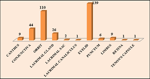
Background: Ophthalmic lesions comprise a wide range of disorders ranging from benign and precancerous to malignant lesions. The rarity of orbito-ocular lesion, further complicates the recognition of their fine and subtle presentations. This can pose a huge diagnostic dilemma to the ophthalmologist and pathologist. This study was undertaken with an intention to study the demographic details, site, clinical presentation and histopathological findings of orbito-ocular lesions to arrive at a definitive diagnosis and for further patient management. Aim: To determine the spectrum of histopathological diagnosis of orbito-ocular lesions. Objectives: 1. To categorize the orbito-ocular lesions into six groups, viz., I-Non-neoplastic (A-Inflammatory, B- Infectious, C- Degenerative, D-Cysts, E-End stage condition, F- Autoimmune disease), II- Precursor lesions, III- Benign, IV- Malignant, V-Metastatic and VI-Syndromic based on the clinical and final histopathological diagnosis.2. To study the frequency distribution of cases according to age, sex, location and clinical presentation of the lesions. 3. To list out the spectrum of orbito-ocular lesions in each category. Materials and Methods: The present study was a retrospective descriptive study conducted for a period of 7 years (2018-2024). Paraffin embedded blocks, slides (including special stains and IHC slides) and previous reports were retrieved and reviewed. Data was collated and analyzed using MS Excel and SPSS software. Results: A total of 348 cases were included in the study. Majority of the cases were in the age group of 51-60 years. Male to female ratio was 1.1:1. Eyelid was the commonest location of lesions constituting 40% (n=139) of the total number of cases. The most common clinical presentation of the lesions was mass (53%, n=186). Non-neoplastic lesions (44.5%, n=155) constituted the majority of cases among all the categories followed by benign lesions (29%, n=102). Among the non-neoplastic lesions, cystic lesions (ID) were predominant constituting 42.5% (n=66) of the total cases. Conclusion: All the ophthalmic biopsies and specimens should be subjected for histopathological examination as the knowledge of spectrum of orbito-ocular lesions will prove to be helpful to the ophthalmologists in shaping the strategy for diagnosis and management of lesions.
##plugins.themes.bootstrap3.article.details##
Copyright (c) 2025 Dr. Seethapathy G, Dr. Suguna B.V., Dr. Shubha H.V., Dr. Vijaya C.

This work is licensed under a Creative Commons Attribution 4.0 International License.
Creative Commons License All articles published in Annals of Medicine and Medical Sciences are licensed under a Creative Commons Attribution 4.0 International License.
Dr. Seethapathy G, Consultant Ophthalmic Plastic and Orbital Surgeon, Manipal hospital, Salem, Tamil Nadu, India.
Consultant Ophthalmic Plastic and Orbital Surgeon, Manipal hospital, Salem, Tamil Nadu, India.
Dr. Suguna B.V., Professor, Department of Pathology, Sapthagiri Institute of Medical Sciences and Research Centre, Bengaluru, Karnataka, India.
Professor, Department of Pathology, Sapthagiri Institute of Medical Sciences and Research Centre, Bengaluru, Karnataka, India.
Dr. Shubha H.V., Associate Professor, Department of Pathology, Sapthagiri Institute of Medical Sciences and Research Centre, Bengaluru, Karnataka, India.
Associate Professor, Department of Pathology, Sapthagiri Institute of Medical Sciences and Research Centre, Bengaluru, Karnataka, India.
Dr. Vijaya C., Professor and HOD, Department of Pathology, Sapthagiri Institute of Medical Sciences and Research Centre, Bengaluru, Karnataka, India.
Professor and HOD, Department of Pathology, Sapthagiri Institute of Medical Sciences and Research Centre, Bengaluru, Karnataka, India.
[1] Ebrahim H, Kedir A, Sena L. Histopathological Patterns of Ophthalmic Lesions and Associated Factors in Jimma University Medical Center, Jimma, South West Ethiopia: a 5-year Retrospective Cross-sectional Study, 2021. Research Square; 2022. DOI: 10.21203/rs.3.rs-1995158/v1.
[2] Bastola, P., Koirala, S., Pokhrel, G., Ghimire, P., &Adhikari, R. (2013). A Clinico-Histopathological Study of Orbital and Ocular Lesions; a Multicenter Study. Journal of Chitwan Medical College, 3(2), 40–44. Retrieved from https://www.nepjol.info/index.php/JCMC/article/view/8442.
[3] Gupta Y, Gahine R, Hussain N, Memon MJ. Clinico- Pathological Spectrum of Ophthalmic Lesions: An Experience in Tertiary Care Hospital of Central India. J Clin Diagn Res. 2017; 11(1): 9-13.
[4] Folberg R. The Eye. In Robbins &Cotran Pathologic Basis of Disease 7th edition, Kumar, V. Abbas, A.K. and Fausto, N (eds). Elsvier Saunders, Philadelphia 2005, pp.1421-1447.
[5] Kayoma DH, Imasogie DE. Histopathological pattern of Orbito-Ocular Lesions, a retrospective hospital-based study spanning 15 years. Afri Health Sci. 2022;22(3): 211-21. https://dx.doi.org/10.4314/ahs.v22i3.23
[6] Rajharsh.D.Hanmante, S.V.Duvernakar, S.A.Deshpande. Histopathological Spectrum of Ophthalmic Lesions: A 5 Year Study. Annals of Pathology and Laboratory Medicine.2018;5(11):A -935-40. DOI: https://doi.org/10.21276/apalm.2101
[7] Gupta Y, Gahine R, Hussain N, Memon MJ. Clinico-Pathological Spectrum of Ophthalmic Lesions: An Experience in Tertiary Care Hospital of Central India. J ClinDiagn Res. 2017 Jan;11(1):EC09-EC13. doi: 10.7860/JCDR/2017/23589.9230. Epub 2017 Jan 1. PMID: 28273971; PMCID: PMC5324416.
[8] Kafle, S. U., Singh, M., Kafle, P. A., KC, B. K., Yadav, S. K., & KC, A. K. (2019). Clinico-histopathological analysis of orbito-ocular lesions: a hospital-based study. Journal of Patan Academy of Health Sciences, 6(2), 39–44. https://doi.org/10.3126/jpahs.v6i2.27228
[9] Paul S, Vo DT, Silkiss RZ. Malignant and benign eyelid lesions in San Francisco: study of a diverse urban population. Am J Clin Med. 2011;8(1):40-6.
[10] Coroi MC, Rosca E, Mutiu G, Coroi T, Bonta M. Eyelid tumours: histopathological and clinical study performed in Country Hospital of Oradea between 2000-2007. Rom J Morphol Embryol Rev. 2010;51(1):111-15.
[11] Kapurdov A, Kapurdova M. Clinical, morphological and epidemiological data on eye tumours. Trakia J of Sciences.2010;8(4):34-9.
[12] Srikanth S. Spectrum of histopathological study of ocular lesions: One year study. J NTR Univ Health Sci. 2014;3(1):12-4. DOI: 10.4103/2277-8632.128417
[13] TikurAnbessa, Menelik. Pattern of ophthalmic lesions at two histopathology centres in Ethiopia. East Afr Med J 2001;78(5):250-4.
[14] Poso M, Mwanza JC, Kayembe DL. Malignant tumours of the eye and adnexa in Congo-Kinshasa. J Fr Ophthalmol 2000;23(4):327-32.