Kimura Disease: A Diagnostic Dilemma in A 51 Years Old Male Presenting with Submandibular Swelling
Authors
##plugins.themes.bootstrap3.article.main##
Abstract
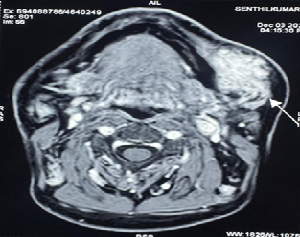
Background: Kimura’s disease is a rare benign chronic inflammatory disorder of unknown origin that involves subcutaneous tissue and lymph nodes. Primarily seen in males predominantly in Asian populations like Chinese and Japanese. The lesion is benign, but it may easily be mistaken for a malignant tumor. Case Presentation: We describe a case of Kimura disease in a 51 years old male presenting with left submandibular swelling. Conclusion: Kimura disease has been confused with angiolymphoid hyperplasia with eosinophilia (ALHE), for which it should be distinguished separately.
##plugins.themes.bootstrap3.article.details##
Copyright (c) 2025 Aniket Kumar, P. Swaminathan, Sukhi R

This work is licensed under a Creative Commons Attribution 4.0 International License.
Creative Commons License All articles published in Annals of Medicine and Medical Sciences are licensed under a Creative Commons Attribution 4.0 International License.
Aniket Kumar, CRRI of Department of Surgery, Karuna Medical College, Palakkad, Kerala, India.
CRRI of Department of Surgery, Karuna Medical College, Palakkad, Kerala, India.
P. Swaminathan, Head of Department of Surgery, Karuna Medical College, Palakkad, Kerala, India.
Head of Department of Surgery, Karuna Medical College, Palakkad, Kerala, India.
Sukhi R, CRRI of Department of Surgery, Karuna Medical College, Palakkad, Kerala, India.
CRRI of Department of Surgery, Karuna Medical College, Palakkad, Kerala, India.
[1] Rao K, Kumar S (2014) Kimura’s Disease-A Rare Cause of Head and Neck Swelling. Int J Otolaryngology Head Neck Surg 3:200-204
[2] Abhange RS, Jadhav RP, Jain NK. Kimura disease: A rare case report. Anl Pathol Lab Med 2018;5:C53 5.
[3] Rajesh A, Prasanth T, Sirisha VN, Azmi MD. Kimura’s disease: A case presentation of postauricular swelling. Niger J Clin Pract 2016; 19:827 30.
[4] Vimal S, Panicker NK, Soraisham P, Chandanwale SS. Kimura disease. Med J Dr. DY Patil Univ 2013; 6:208.
[5] Hui PK, Chan JK, Ng CS, Kung IT, Gwi E. Lymphadenopathy of Kimura’s disease. Am J Surg Pathol. 1989; 13:177-186.
[6] Kini U, Shariff S. Cytodiagnosis of Kimura’s disease. Indian J Pathol Microbiol. 1998; 41:473-477.
[7] Abhay H, Swapna S, Darshan T, Vishal J, Gautam P. Kimura’s Disease: A Rare Cause of Local Lymphadenopathy. Int J Sci Stud. 2014; 2:122-125.
[8] Meningaud JP, Pitak-Arnnop P, Fouret P, Bertrand JC. Kimura’s disease of the parotid region: report of 2 cases and review of the literature. J Oral Maxillofac Surg. 2007; 65:134-140.
[9] Chen H, Thompson LD, Aguilera NS, Abbondanzo SL. Kimura disease: a clinicopathologic study of 21 cases. Am J Surg Pathol. 2004; 28:505-513.
[10] Tseng CF, Lin HC, Huang SC, Su YC. Kimura’s disease presenting as bilateral parotid masses. Eur Arch of Otorhinolaryngol. 2005; 262:8-10.
[11] Kimura T, Yoshimura S, Ishikaura E. Unusual granulation combined with hyperplastic changes of lymphatic tissue. Trans Soc Pathol Jpn. 1948; 37:179-180.
[12] Kim BH, Sithian N, Cucolo GF. Subcutaneous angiolymphoid hyperplasia (Kimura disease). Report of a case. Arch Surg.1975;110 :1246– 1248.

