
A Case Control Study on Histomorphological Changes of Fallopian Tube in Surface Epithelial Tumors of Ovary at a Tertiary Care Center
Authors

##plugins.themes.bootstrap3.article.sidebar##
##plugins.themes.bootstrap3.article.main##
Abstract
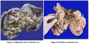
Background: Ovarian cancer, specifically high-grade serous carcinoma, is among the top causes of cancer-related mortality in women, and there is no reliable screening test for its early detection. New evidence indicates that the fallopian tube, and more precisely its fimbrial part, may be the origin of most ovarian serous carcinomas, thereby going against the conventional belief placing the site of origin in the ovarian surface epithelium. Aim and objective: The aim of this research was to examine the relationship of histomorphological changes in the epithelium of the fallopian tubes with ovarian surface epithelial tumors. The main objective was to compare the epithelial changes in the Fallopian tube from panhysterectomy specimen of cases with different types of surface epithelial ovarian tumors (benign, borderline, or malignant) and cases with benign uterine lesions. Material and Methods: A case-control study was done during November 2020 to November 2021 at Lakeshore Hospital and Research Center, Kochi. Forty-eight patients were recruited: 24 cases of epithelial ovarian tumors (10 benign, 5 borderline, 9 malignant) and 24 controls of benign uterine lesions. Fallopian tubes from panhysterectomy specimens were graded for tubal involvement, epithelial stratification, atypia, mitosis, intraepithelial vacuoles, and other changes histologically. Statistical significance was determined using a p-value cut-off of 0.05. Results: The case group had significantly greater frequencies of epithelial stratification (37.5% vs. 4.2%, p<0.05), atypia (12.5% vs. 0.0%, p<0.05), mitosis (20.8% vs. 0.0%, p<0.05), and intraepithelial vacuoles (12.5% vs. 0.0%, p<0.05) compared with the control group. Tube involvement occurred in 4 of 9 malignant cases (44.4%), and serous tubal intraepithelial carcinoma (STIC) occurred in one high-grade serous carcinoma case (11.1%). No secretory cell outgrowth occurred in either group. Conclusion: The research establishes a significant correlation between histomorphological changes in the epithelium of the fallopian tube and ovarian serous carcinomas and thereby supports the postulate that the fallopian tube (fimbrial end) is a potential site of origin of the neoplasms. The research emphasizes the importance of examining fallopian tubes in the context of ovarian cancer and has preventive surgical implications such as salpingectomy.
##plugins.themes.bootstrap3.article.details##
Copyright (c) 2025 Shahseena Abdulla, Iona Leekha Mathew, Rosmy John, Chithrathara K, Pushpa Mahadevan, Ami Maria Emmanuel

This work is licensed under a Creative Commons Attribution 4.0 International License.
Creative Commons License All articles published in Annals of Medicine and Medical Sciences are licensed under a Creative Commons Attribution 4.0 International License.
Iona Leekha Mathew, Department of Pathology, Karuna Medical College, Vilayodi, Chittur, Palakkad, Kerala, 678103, India.
Department of Pathology, Lakeshore Hospital and Research Centre, Kochi, Kerala, INDIA
[1] Hohn AK, Brambs CE, Hiller GGR, May D, Schmoeckel E, Horn LC. 2020 WHO Classification of Female Genital Tumors. Geburtshilfe Frauenheilkd. 2021 Oct;81(10):1145-1153. doi: 10.1055/a-1545-4279. Epub 2021 Oct 6. PMID: 34629493; PMCID: PMC8494521.
[2] Dubeau L. The cell of origin of ovarian epithelial tumours. Lancet Oncol 2008; 9:1191–7. [PubMed: 19038766]
[3] Diniz PM, Carvalho JP, Baracat EC, Carvalho FM. Fallopian tube origin of supposed ovarian high grade serous carcinomas. Clinics (Sao Paulo) 2011; 66:73 6
[4] Crum CP, Drapkin R, Miron A, Ince TA, Muto M, Kindelberger DW, et al. The distal fallopian tube: A new model for pelvic serous carcinogenesis. Curr Opin Obstet Gynecol 2007; 19:3 9
[5] Vang R, Shih IM, Kurman RJ. Ovarian low grade and high-grade serous carcinoma: Pathogenesis, clinicopathologic and molecular biologic features, and diagnostic problems. Adv Anat Pathol 2009; 16:267 82
[6] Liang Y, Chen XD, Lü BJ, Zhou CY, Zhang XF, Shi HY. Preliminary study on the relationship between tubal intraepithelial carcinoma of the fimbria and pelvic high grade serous carcinoma. Zhonghua Fu Chan Ke Za Zhi 2011; 46:724 8
[7] Crum CP. Intercepting pelvic cancer in the distal fallopian tube: Theories and realities. Mol Oncol 2009; 3:165 70.
[8] Jarboe E, Folkins A, Nucci MR, Kindelberger D, Drapkin R, Miron A, et al. Serous carcinogenesis in the fallopian tube: A descriptive classification. Int J Gynecol Pathol 2008; 27:1 9
[9] Sehdev AS, Kurman RJ, Kuhn E, Shih IM. Serous tubal intraepithelial carcinoma upregulates markers associated with high grade serous carcinomas including Rsf 1 (HBXAP), cyclin E and fatty acid synthase. Mod Pathol 2010; 23:844 55
[10] Gilks CB, Prat J. Ovarian carcinoma pathology and genetics: Recent advances. Hum Pathol 2009; 40:1213 23
[11] Kobayashi H, Iwai K, Niiro E, Morioka S, Yamada Y, Ogawa K, et al. The conceptual advances of carcinogenic sequence model in high grade serous ovarian cancer. Biomed Rep 2017; 7:209 13
[12] Neel BG, Zhang S, Zhang T, Dolgalev I, Ran H, Levine DA. Both Fallopian Tube and Ovarian Surface Epithelium Can Act as Cell of Origin for High Grade Serous Ovarian Carcinoma; 2018. p. 481200
[13] Callahan MJ, Crum CP, Medeiros F, et al. Primary fallopian tube malignancies in BRCApositive women undergoing surgery for ovarian cancer risk reduction. J Clin Oncol 2007;25: 3985-90.
[14] Cass I, Holschneider C, Datta N, et al. BRCA-mutation-associated fallopian tube www.AJOG.org Oncology Expert Reviews carcinoma: a distinct clinical phenotype? Obstet Gynecol 2005;106:1327-34.
[15] Leeper K, Garcia R, Swisher E, et al. Pathologic findings in prophylactic oophorectomy specimens in high-risk women. Gynecol Oncol 2002;87:52-6
[16] Piek JM, van Diest PJ, Zweemer RP, et al. Dysplastic changes in prophylactically removed fallopian tubes of women predisposed to developing ovarian cancer. J Pathol 2001;195: 451-6
[17] Shaw PA, Rouzbahman M, Pizer ES, Pintilie M, Begley H. Candidate serous cancer precursors in fallopian tube epithelium of BRCA1/2 mutation carriers. Mod Pathol. 2009; 22: 1133–1138
[18] Kurman RJ, Shih IM. Molecular pathogenesis and extraovarian origin of epithelial ovarian cancer – Shifting the paradigm. Hum Pathol 2011; 42:918 31 Histol Histopathol, Vol 21, Scott and McCluggage. [cited 2019 Oct 20].
[19] Semmel DR, Folkins AK, Hirsch MS, Nucci MR, Crum CP. Intercepting early pelvic serous carcinoma by routine pathological examination of the fimbria. Mod Pathol.2009;22:985-988.
[20] Bergsten TM, Burdette JE, Dean M. Fallopian tube initiation of high grade serous ovarian cancer and ovarian metastasis: Mechanisms and therapeutic implications. Cancer letters. 2020 Apr 28; 476:152-60.
[21] Aslani FS, Maleknasab M, Akbarzadeh-Jahromi M. Fallopian tube epithelial changes in ovarian serous tumors compared with control group: A single-center study. Nigerian medical journal: journal of the Nigeria Medical Association. 2019 Mar;60(2):47.
[22] Vang R, Shih I-M, Kurman RJ. Ovarian Low-grade and High-grade Serous Carcinoma: Pathogenesis, Clinicopathologic and Molecular Biologic Features, and Diagnostic Problems. Adv Anat Pathol. 2009 Sep;16(5):267-82.
[23] Hunt JL, Lynn AA. Histologic features of surgically removed fallopian tubes. Arch Pathol Lab Med 2002; 126:951 5.
[24] Li J, Fadare O, Xiang L, Kong B, Zheng W. Ovarian serous carcinoma: Recent concepts on its origin and carcinogenesis. J Hematol Oncol 2012; 5:8.
[25] Hankinson SE, Hunter DJ, Colditz GA, Willett WC, Stampfer MJ, Rosner B, Hennekens CH, et al. Tubal ligation, hysterectomy, and risk of ovarian cancer. A prospective study. JAMA 1993; 270: 2813-2818.