
Profile and Prognostic Value of Mucocutaneous Lesions in Children Hospitalized for Severe Acute Malnutrition in Nouakchott, Mauritania
Authors

##plugins.themes.bootstrap3.article.sidebar##
##plugins.themes.bootstrap3.article.main##
Abstract
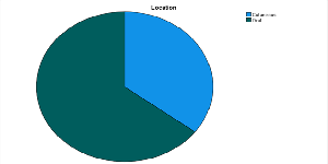
Introduction: Severe acute malnutrition (SAM) remains a major public health problem and an important cause of morbidity and mortality among children in resource-limited settings. Cutaneous and mucosal lesions are frequent clinical manifestations in children with SAM and result from multiple nutritional deficiencies and associated infections. These lesions may serve as early and visible markers of disease severity and poor prognosis. Objective: To describe the profile of mucocutaneous lesions in children hospitalized for Severe acute malnutrition and to analyze their prognostic value in relation to clinical complications. Methods: A retrospective descriptive and analytical study was conducted at the Inpatient Nutritional Rehabilitation and Education Center of the National Hospital Center in Nouakchott. Medical records of children under 59 months hospitalized for Severe acute malnutrition were reviewed. Associations between mucocutaneous lesions, sociodemographic variables, and clinical complications were analyzed using the chi-square test or Fisher’s exact test (p < 0.05). Results: A total of 147 children were included, with a mean age of 14.6 months; 56.5% were boys. The prevalence of mucocutaneous lesions was 23.1%, predominantly oral (64.7%), mainly oral thrush (29.5%), aphthous ulcers (17.6%), and angular cheilitis (17.6%). No significant association was found with age, sex, or place of origin, whereas a significant association was observed with the quarter of hospitalization (p = 0.01). Children with lesions more frequently presented multiple complications, including anemia, cough, and dehydration, with a significant association with more than two concomitant complications (p = 0.01). Conclusion: Mucocutaneous lesions are common in children with Severe acute malnutrition and may serve as markers of increased morbidity, highlighting the importance of systematic screening at admission.
##plugins.themes.bootstrap3.article.details##
Copyright (c) 2026 Mariem Sidi Mohamed, Aicha Biha, Mohmed Aly Lemrabott, Setty Sass, Ahmed Salem Cheikh Baba, Lella Abdellahi Hamedy, Ahmed El Baraa, Ahmed Feil

This work is licensed under a Creative Commons Attribution 4.0 International License.
Creative Commons License All articles published in Annals of Medicine and Medical Sciences are licensed under a Creative Commons Attribution 4.0 International License.
[1] Onasaka LS, Makivovela DM, Kabeya CM. Profile of severe acute malnutrition in children aged 6–59 months in the Intensive Therapeutic Nutrition Unit of the Kinshasa General Reference Hospital, DRC. Int J Prog Sci Technol 2022; 2: 1–7.
[2] Ministry of Health of Burkina Faso. National protocol for the integrated management of acute malnutrition (IMAM). Ouagadougou: Ministry of Health; 2014: 1–12.
[3] Garenne M, Maire B, Fontaine O, Dieng K, Briend A. A criterion for the prevalence of malnutrition: child survival. In: Nutritional Deficiencies in Developing Countries. Paris: Karthala; 1995: 12–19.
[4] Schaible UE, Kaufmann SHE. Malnutrition and infection: complex mechanisms and global impacts. PLoS Med 2007; 4: e115. doi:10.1371/journal.pmed.0040115
[5] Rytter MJH, Kolte L, Briend A, Friis H, Christensen VB. The immune system in children with malnutrition: a systematic review. PLoS One 2014; 9: e105017. doi:10.1371/journal.pone.0105017
[6] Marelli A, D’Hollander K, de Polnay K. Newborns, children with severe malnutrition and children <5 years with major wounds. Brussels: Médecins Sans Frontières; 2019: 1–9.
[7] Adégbidi H, Degboé B, Saka B, Elégbédé A, Atadokpèdé F, Koudoukpo C, et al. Profile of immune and allergic dermatoses among children at the outpatient dermatology clinic in Cotonou (Benin). Med Sante Trop 2014; 24: 446–448. doi:10.1684/mst.2014.0405
[8] Diabaté A, Kourouma S, Kouabenan AA, Gué I, Vagamon B, Aka BR. Epidemiological, clinical and evolutionary profile of superficial cutaneous parasitic infections in hospitals in Ivory Coast. Rev Int Sci Med Abidjan 2018; 20: 67–70.
[9] Fofana Y, Traoré B, Dicko A, Faye O, Berthe S, Cisse L, et al. Epidemio-clinical profile of dermatoses in children receiving dermatological consultation in Bamako (Mali). Pan Afr Med J 2016; 25: 238. doi:10.11604/pamj.2016.25.238.10564
[10] Somé N, Zoungrana A, Konaté I, Ouédraogo M, Tapsoba GP, Sosso-Kargougou N, et al. Skin disorders in preschool environment in the city of Ouagadougou (Burkina Faso). Our Dermatol Online 2019; 10: e31. doi:10.7241/ourd.2019e.31
[11] Tchangaï-Walla K, Pitché P, Agbèrè A, Bakondé B. Reasons for children's dermatology consultations in Lomé (Togo). Med Afr Noire 1995; 42: 390–392.
[12] Keita M. Epidemiological and clinical study of dermatoses in children aged 0–15 years at CNUAM Bamako [Doctoral thesis]. Bamako: Faculty of Medicine and Odontology-Stomatology; 2013: 1–14.
[13] World Health Organization, United Nations Children’s Fund. WHO child growth standards and the identification of severe acute malnutrition in infants and children: a joint statement. Geneva: WHO; 2009.
[14] Crouma K. Child nutrition care at the reference health center of commune V of Bamako [Thesis]. Bamako: University of Bamako, Faculty of Medicine, Pharmacy and Odontostomatology; 2008: 1–83.
[15] Issa D. Evaluation of the management of severe acute malnutrition in children aged 6–59 months at the URENI of Koutiala [Thesis]. Koutiala: Faculty of Medicine and Odontostomatology; 2015: 1–21.
[16] Diarra S. Study of dermatoses in infants consulting in the dermatology department of the CNAM [Doctoral thesis]. Bamako: Faculty of Medicine and Odontostomatology; 2015: 38–54.
[17] Aminata F. Determining factors in marasmus and kwashiorkor in children aged 6–59 months at URENI Kalaban Coro [Thesis]. Bamako: University of Sciences, Techniques and Technologies of Bamako; 2021: 1–14.
[18] Aimé B, Mukuku O, Kasongo K, et al. Clinical signs encountered in malnourished children in a mining environment: Lubumbashi and surroundings. Pan Afr Med J 2016; 24: 67.
[19] Moctar M. Dermatological manifestations in severely malnourished children in Niamey hospitals [Thesis]. Niamey: Faculty of Agronomy; 2018: 12–25.
[20] Vishalakshi S, Pandit KU. A cross-sectional study of nutritional dermatoses among malnourished children in a tertiary care centre. Indian J Paediatr Dermatol 2021; 22: 226–230. doi:10.4103/ijpd.IJPD_13_20
[21] Action contre la Faim. Malnutrition: un fléau qui pourrait toucher 2 milliards de personnes. Paris: Action contre la Faim; 2020.