
Diagnostic Accuracy of Magnetic Resonance Imaging in Assessing Myometrial Invasion in Endometrial Carcinoma: A Retrospective Single Centre Study
Authors

##plugins.themes.bootstrap3.article.sidebar##
##plugins.themes.bootstrap3.article.main##
Abstract
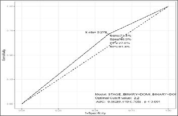
Background: Endometrial carcinoma is the most common gynecologic malignancy in developed nations. Accurate preoperative assessment of myometrial invasion is critical for staging and guiding surgical management. This study aimed to evaluate the diagnostic accuracy of 1.5 Tesla plain magnetic resonance imaging (MRI) in determining the depth of myometrial invasion compared with postoperative histopathology. Materials and Methods: A retrospective analysis was conducted on 189 patients with histologically confirmed endometrial carcinoma who underwent preoperative 1.5 T plain MRI using T1- and T2-weighted sequences. MRI findings were correlated with histopathological staging, and diagnostic indices, including sensitivity, specificity, predictive values, and area under the curve (AUC), were calculated across FIGO stages. Results: The mean patient age was 56.3 years, predominantly postmenopausal. MRI accuracy for detecting myometrial invasion was 85.8 % for Stage IA, 63.8 % for Stage IB, and 66.6 % for Stage II disease. Overall, MRI demonstrated 27.8 % sensitivity, 81.8 % specificity, 71.4 % PPV, and 40.9 % NPV, with an AUC of 0.562. Conclusion: Plain 1.5 T MRI showed moderate diagnostic accuracy with high specificity for early-stage endometrial carcinoma but limited sensitivity for advanced invasion. The study underscores MRI’s role as a practical preoperative tool, particularly in resource-limited settings lacking contrast or diffusion-weighted imaging.
##plugins.themes.bootstrap3.article.details##
Copyright (c) 2025 S. Subbiah, J. Sakthi Usha Devi, S. Karnan Srinivas, S. Dorian Hanniel Terrence, Sasikala Kathiresan, Emil Phinehas Mariantony

This work is licensed under a Creative Commons Attribution 4.0 International License.
Creative Commons License All articles published in Annals of Medicine and Medical Sciences are licensed under a Creative Commons Attribution 4.0 International License.
S. Subbiah, Professor and HOD, Department of Surgical Oncology, Govt. Arignar Anna Memorial Cancer Hospital and Research Institute, Karapettai, Kanchipuram, Tamil Nadu, India
Professor and HOD, Department of Surgical Oncology, Govt. Arignar Anna Memorial Cancer Hospital and Research Institute, Karapettai, Kanchipuram, Tamil Nadu, India
J. Sakthi Usha Devi, Associate Professor, Department of Surgical Oncology, Kalaignar Centenary Super-Speciality Hospital, Guindy, Chennai, Tamil Nadu, India.
Associate Professor, Department of Surgical Oncology, Kalaignar Centenary Super-Speciality Hospital, Guindy, Chennai, Tamil Nadu, India.
S. Karnan Srinivas, Assistant Professor, Department of Surgical Oncology, Govt. Arignar Anna Memorial Cancer Hospital and Research Institute, Karapettai, Kanchipuram, Tamil Nadu, India.
Assistant Professor, Department of Surgical Oncology, Govt. Arignar Anna Memorial Cancer Hospital and Research Institute, Karapettai, Kanchipuram, Tamil Nadu, India.
S. Dorian Hanniel Terrence, Senior Resident, Department of Surgical Oncology, Govt. Arignar Anna Memorial Cancer Hospital and Research Institute, Karapettai, Kanchipuram, Tamil Nadu, India.
Senior Resident, Department of Surgical Oncology, Govt. Arignar Anna Memorial Cancer Hospital and Research Institute, Karapettai, Kanchipuram, Tamil Nadu, India.
Sasikala Kathiresan, Assistant Professor Department of Obstetrics and Gynecology, AIIMS, Madurai, Tamil Nadu, India.
Assistant Professor Department of Obstetrics and Gynecology, AIIMS, Madurai, Tamil Nadu, India.
Emil Phinehas Mariantony, Assistant Professor, Department of General Surgery, AIIMS, Madurai, Tamil Nadu, India.
Assistant Professor, Department of General Surgery, AIIMS, Madurai, Tamil Nadu, India.
[1] Mahdy H, Vadakekut ES, Crotzer D. Endometrial Cancer. [Updated 2024 Apr 20]. In: StatPearls [Internet]. Treasure Island (FL): StatPearls Publishing; 2025 Jan-. Available from: https://www.ncbi.nlm.nih.gov/books/NBK525981/
[2] Tang, W. Z., Cai, Q. Y., Huang, K. J., et al. (2025). The global burden of polycystic ovary syndrome, endometriosis, uterine fibroids, cervical cancer, uterine cancer, and ovarian cancer from 1990 to 2021. BMC Public Health, 25(1), 1774.
[3] Okuda, T., Sekizawa, A., Purwosunu, Y., et al. (2010). Genetics of endometrial cancers. Obstetrics and gynecology international, 2010, 984013. https://doi.org/10.1155/2010/984013
[4] Espinosa, I., D'Angelo, E., & Prat, J. (2024). Endometrial carcinoma: 10 years of TCGA (the cancer genome atlas): A critical reappraisal with comments on FIGO 2023 staging. Gynecologic Oncology, 186, 94-103.
[5] Pinto, A. P. (2025). The role of ultrasound in preoperative staging in patients with advanced ovarian cancer.
[6] Maheshwari, E., Nougaret, S., Stein, et al. (2022). Update on MRI in evaluation and treatment of endometrial cancer. Radiographics, 42(7), 2112-2130.
[7] Jin, X., Shen, C., Yang, X., et al. (2022). Association of tumor size with myometrial invasion, lymphovascular space invasion, lymph node metastasis, and recurrence in endometrial cancer: a meta-analysis of 40 studies with 53,276 patients. Frontiers in oncology, 12, 881850.
[8] Nougaret, S., Sala, E., Lakhman, Y., et al. (2025). Updated ESUR Guidelines for Endometrial Cancer: integrating MRI with the 2023 FIGO Staging Revolution. European Radiology, 1-16.
[9] Takeuchi, M., Matsuzaki, K., & Harada, M. (2018). Evaluating myometrial invasion in endometrial cancer: comparison of reduced field-of-view diffusion-weighted imaging and dynamic contrast-enhanced MR imaging. Magnetic Resonance in Medical Sciences, 17(1), 28-34.
[10] Zamani, F., Goodarzi, S., Hallaji, F., et al. (2012). Diagnostic Value of Pelvic MRI for Assessment of the Depth of Myometrial Invasion and Cervical Involvement in Endometrial Cancer: Comparison of New Versus Old FIGO Staging. Iranian journal of radiology: a quarterly journal published by the Iranian Radiological Society, 9(4), 202–208. https://doi.org/10.5812/iranjradiol.5276
[11] Gul, P., Gul, K., Altaf, M. O., et al. (2022). The accuracy of MRI in the local staging of endometrial cancer: an experience from a tertiary care oncology Institute in Pakistan. Cureus, 14(11).
[12] Sanjuán, A., Escaramís, G., Ayuso, J. R., et al. (2008). Role of magnetic resonance imaging and cause of pitfalls in detecting myometrial invasion and cervical involvement in endometrial cancer. Archives of gynecology and obstetrics, 278(6), 535–539. https://doi.org/10.1007/s00404-008-0636-1
[13] Sajid, A., Wahid, G., Jawaid, A., et al. (2025). DIAGNOSTIC ACCURACY OF DIFFUSION-WEIGHTED MAGNETIC RESONANCE IMAGING IN DETECTING DEEP MYOMETRIAL INVASION BY ENDOMETRIAL TUMOR TAKING HISTOPATHOLOGY AS GOLD STANDARD. Khyber Journal of Medical Sciences, 18(3), 243-248.
[14] Akçay, A., & Peker, A. A. (2025). Diagnostic Comparison of MRI Sequences for Assessing Myometrial Invasion in Endometrial Cancer: A 1.5 T MRI Study. Hitit Medical Journal, 7(1), 29-36.