
Clinical Significance and Variations of Fetal Frontomaxillary Facial Angle Measurement in North Indian Pregnant Females
Authors

##plugins.themes.bootstrap3.article.sidebar##
##plugins.themes.bootstrap3.article.main##
Abstract
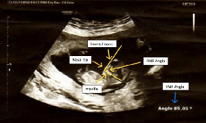
Objective: This study aims to establish normative reference ranges for FMF angle measurements in North Indian fetuses during first trimester (11-13+6 weeks of gestation) and assess its clinical significance. Design: A prospective observational study was conducted at GSVM Medical College, Kanpur, involving 161 North Indian pregnant females. Methods: Transabdominal ultrasound measurements of FMF angle were obtained using standardized mid-sagittal imaging techniques. The relationship between FMF angle and crown-rump length (CRL), gestational age, and maternal factors was analyzed using statistical models. Results: The mean FMF angle was 83.90° (SD = 1.76°), with a range of 80.0° to 87.3°. A strong inverse correlation was observed between FMF angle and CRL (r = -0.789, p <.001), confirming a gestational age-dependent decline. No significant influence of maternal age (p =.574) or gravida status (p =.233) was found on FMF angle measurements. The 5th to 95th percentile reference range for FMF angle was established as 81.3° to 86.5°, providing a clinically relevant baseline for North Indian populations. Conclusion: FMF angle is a stable and reproducible prenatal screening parameter in North Indian pregnancies. The study establishes normative data essential for refining regional first- trimester screening protocols and improving early detection of fetal anomalies.
##plugins.themes.bootstrap3.article.details##
Copyright (c) 2025 Ashok K. Verma, Vannekuti Vinay, Pavika Lal, Renu Gupta, Poonam S. Gambhir, Garima Gupta, Deepak Anand

This work is licensed under a Creative Commons Attribution 4.0 International License.
Creative Commons License All articles published in Annals of Medicine and Medical Sciences are licensed under a Creative Commons Attribution 4.0 International License.
Ashok K. Verma, Associate Professor, Department of Radiodiagnosis, GSVM Medical College, Kanpur, India.
Associate Professor, Department of Radiodiagnosis, GSVM Medical College, Kanpur, India.
Vannekuti Vinay, Associate Professor, Department of Obstetrics and Gynaecology, GSVM Medical College, Kanpur, India.
Associate Professor, Department of Obstetrics and Gynaecology, GSVM Medical College, Kanpur, India.
Pavika Lal, Associate Professor, Department of Obstetrics and Gynaecology, GSVM Medical College, Kanpur, India.
Associate Professor, Department of Obstetrics and Gynaecology, GSVM Medical College, Kanpur, India.
Renu Gupta, Associate Professor, Department of Obstetrics and Gynaecology, GSVM Medical College, Kanpur, India.
Associate Professor, Department of Obstetrics and Gynaecology, GSVM Medical College, Kanpur, India.
Poonam S. Gambhir, Consultant Clinical Genetics and Fetal Medicine at Vardaan Genetic and Diagnostic Centre, Kanpur, India.
Consultant Clinical Genetics and Fetal Medicine at Vardaan Genetic and Diagnostic Centre, Kanpur, India.
Garima Gupta, Associate Professor, Department of Obstetrics and Gynaecology, GSVM Medical College, Kanpur, India.
Associate Professor, Department of Obstetrics and Gynaecology, GSVM Medical College, Kanpur, India.
Deepak Anand, Associate Professor, Department of Obstetrics and Gynaecology, GSVM Medical College, Kanpur, India.
Associate Professor, Department of Obstetrics and Gynaecology, GSVM Medical College, Kanpur, India.
[1] Mangla, Mishu; Kumar, Naina: First Trimester Ultrasound Soft Markers in a Fetus: Genetic Associations and Diagnostic Implications. Maternal-Fetal Medicine. 2025, 40:1-12. 10.1097/FM9.0000000000000301
[2] Raniga S, Desai PD, Parikh H.: Ultrasonographic soft markers of aneuploidy in second trimester: are we lost?. MedGenMed. 2006, 8(1):9:8(1):9.
[3] Jiri Sonek, Marisa Borenstein, Themistoklis Dagklis, Nicola Persico, Kypros H. Nicolaides,: Frontomaxillary facial angle in fetuses with trisomy 21 at 11-13(6) weeks. Am J Obstet Gynecol. 2007, 196:10.1016/j.ajog.2006.10.891
[4] S Pranpanus 1, O Kor-anantakul, T Suntharasaj, C Suwanrath, R Leetanaporn, T Hanprasertpong, N Pruksanusak.: Frontomaxillary facial angle in chromosomally normal Thai foetuses at 11 to 13 weeks 6 days ’ gestation. J Obstet Gynaecol. 2016, 36:53-7. 10.3109/01443615.2015.1030598.
[5] Alphonse J, Cox J, Clarke J, Schluter P, McLennan A: The Effect of Ethnicity on 2D and 3D Frontomaxillary Facial Angle Measurement in the First Trimester. Obstet Gynecol Int. 2013, 2013:10.1155/2013/847293
[6] W. Plasencia, T. Dagklis, A. Sotiriadis, M. Borenstein, K. H. Nicolaides: Frontomaxillary facial angle at 11 + 0 to 13 + 6 weeks' gestation—reproducibility of measurements. Ultrasound in obstetrics & gynecology. 2006, 29:18-21. 10.1002/uog.3907
[7] Czuba B, Cnota W, Wloch A, Wegrzyn P, Sodowski K, Wielgos M, Borowski D: Frontomaxillary facial angle measurement in screening for trisomy 18 at 11 + 0 to 13 + 6 weeks of pregnancy: a double-centre study. Biomed Res Int. 2013, 2013:1-4. 10.1155/2013/168302
[8] M Borenstein, N Persico, T Dagklis, E Faros, K H Nicolaides: Frontomaxillary facial angle in fetuses with trisomy 13 at 11 + 0 to 13 + 6 weeks. Ultrasound in obstetrics & gynecology. 2007, 30:818-823. 10.1002/uog.5135
[9] R. Lachmann, G. Picciarelli, J. Moratalla, N. Greene, K. H. Nicolaides: Frontomaxillary facial angle in fetuses with spina bifida at 11-13 weeks' gestation. Ultrasound in obstetrics & gynecology. 2010, 36:268-271. 10.1002/uog.7718
[10] C. Hsiao: Frontomaxillary facial angle in the normal and trisomy 21 at 11 and 13 + 6 weeks gestation in Asian population. Ultrasound in obstetrics & gynecology. 2011, 38:186. 10.1002/uog.9678
[11] F Molina, N Persico, M Borenstein, J Sonek, K H Nicolaides: Frontomaxillary facial angle in trisomy 21 fetuses at 16-24 weeks of gestation. Ultrasound in obstetrics & gynecology. 2008, 31:384-387. 10.1002/uog.5288
[12] M. BORENSTEIN, N. PERSICO, I. STROBL, J. SONEK and K. H. NICOLAIDES: Frontomaxillary and mandibulomaxillary facial angles at 11 + 0 to 13 + 6 weeks in fetuses with trisomy 18. Ultrasound in obstetrics & gynecology. 2007, 30: 928-933. 10.1002/uog.5188
[13] Roshan Jabeen, Karthika P: Updated socioeconomic classification: revised modified B. G. Prasad and modified Kuppuswamy scales for January 2025. International Journal of Community Medicine and Public Health. 2025, 12:2103. 10.18203/2394-6040.ijcmph20251038
[14] C. H. Hsiao, C. L. Lin, C. F. Hsieh: Frontomaxillary facial angle in normal fetuses at 11 + 0 to 13 + 6 weeks in an oriental population. Ultrasound in obstetrics & gynecology. 2008, 32:310. 10.1002/uog.5613
[15] Ana Paula Nascimento Panigassi, Edward Araujo Júnior, Luciano Marcondes Machado Nardozza, Antonio Fernandes Moron & David Baptista da Silva Pares: Fetal frontomaxillary facial angle between 11 and 13 + 6 weeks of gestation in a Brazilian population: influence of different races. The Journal of Maternal-Fetal & Neonatal Medicine. 2013, 26: 1116-1120. 10.3109/14767058.2013.771164
[16] Jakub Staniczek, Maisa Manasar-Dyrbuś, Magda Rybak-Krzyszkowska, Adrianna Kondracka, Dominika Orszulak: Systematic review and meta-analysis of the association between young maternal age and fetal abnormalities. Sci Rep. 2024, 14:10.1038/s41598-024-74015-1