
Microbiology of Pin Site Infections in a Nigerian Tertiary Hospital: A Prospective Analysis
Authors

##plugins.themes.bootstrap3.article.sidebar##
##plugins.themes.bootstrap3.article.main##
Abstract
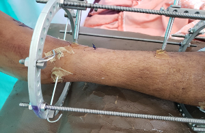
Background: Pin site infection remains the most common complication following external fixation, with reported incidence rates varying widely. Understanding the microbiological profile of these infections is crucial for developing effective prevention and treatment strategies in resource-limited settings. Objective: To determine the microbiological profile, bacterial load, and infection rates of pin sites in patients undergoing external fixation at a Nigerian tertiary hospital, comparing two dressing protocols. Methods: A randomized controlled trial was conducted over 10 months involving 50 patients undergoing external fixation at the University College Hospital, Ibadan. Patients were randomized into two groups: Medi-Honey™ dressing (n=25) and povidone-iodine gauze dressing (n=25). Pin site cultures were obtained on the 5th postoperative day for microscopy, culture, and bacterial count. Clinical monitoring for infection was performed weekly for 4 weeks using the Dahl Wire Pin Site Classification system. Results: A total of 328 pin sites were evaluated, with an overall infection rate of 7.01% (23/328). The infection rate was significantly lower in the Medi-Honey™ group (3.70%) compared to the povidone-iodine group (11.51%) (p=0.001, RR=0.32, 95%CI: 0.14-0.76). Staphylococcus aureus was the predominant organism cultured (71.4%), followed by Staphylococcus epidermidis (28.6%). All cultured organisms were gram-positive cocci. Bacterial counts ranged from 58 to 126 CFU/ml. No significant association was found between bacteriological parameters and clinical outcomes (p=0.42). Conclusion: The study demonstrates a relatively low pin site infection rate with gram-positive cocci as the predominant pathogens. Medi-Honey™ dressing showed superior antimicrobial efficacy compared to povidone-iodine. These findings support the need for targeted antimicrobial strategies against staphylococcal species in pin site care protocols.
##plugins.themes.bootstrap3.article.details##
Copyright (c) 2025 Adeoye Allen-Taylor, Aliu Olalekan Olatunji, Jemiludeen O. Morhason-Bello, Michael O. Okunola, Mosimabale J. Balogun

This work is licensed under a Creative Commons Attribution 4.0 International License.
Creative Commons License All articles published in Annals of Medicine and Medical Sciences are licensed under a Creative Commons Attribution 4.0 International License.
Adeoye Allen-Taylor, Department of Orthopaedics and Trauma, University College Hospital, Ibadan, Oyo State, Nigeria.
Department of Orthopaedics and Trauma, University College Hospital, Ibadan, Oyo State, Nigeria.
Aliu Olalekan Olatunji, Department of Medical Microbiology, University College Hospital, Ibadan, Oyo State, Nigeria.
Department of Medical Microbiology, University College Hospital, Ibadan, Oyo State, Nigeria.
Jemiludeen O. Morhason-Bello, Department of Orthopaedics and Trauma, University College Hospital, Ibadan, Oyo State, Nigeria.
Department of Orthopaedics and Trauma, University College Hospital, Ibadan, Oyo State, Nigeria.
Michael O. Okunola, Department of Orthopaedics and Trauma, University College Hospital, Ibadan, Oyo State, Nigeria.
Department of Orthopaedics and Trauma, University College Hospital, Ibadan, Oyo State, Nigeria.
Mosimabale J. Balogun, Department of Surgery, College of Medicine, University of Ibadan, Oyo State, Nigeria.
Department of Surgery, College of Medicine, University of Ibadan, Oyo State, Nigeria.
[1] Pape HC, Tornetta P, Tarkin I, Tzioupis C, Sabeson V, Olson SA. Timing of fracture fixation in multitrauma patients: the role of early total care and damage control surgery. J Am Acad Orthop Surg. 2009;17(9):541-9.
[2] Marin LE, McBroom DB, Caban G. Percutaneous reduction and external fixation for foot and ankle fractures. Clin Podiatr Med Surg. 2008;25(4):721-32.
[3] Pfahler M, Krodel A, Tritschler A, Zenta S. Role of internal and external fixation in ankle fusion. Arch Orthop Trauma Surg. 1996;115(3-4):146-8.
[4] Handelsman JE, Weinberg J, Razi A, Mulley DA. The role of AO external fixation in proximal femoral osteotomies in the pediatric neuromuscular population. J Pediatr Orthop Part B. 2004;13(5):303-7.
[5] Bini A, Surace MF, Pilato G. Complex articular fractures of the distal radius: the role of closed reduction and external fixation. J Hand Surg Eur Vol. 2008;33(3):305-10.
[6] Osman W, Alaya Z, Kaziz H, et al. Treatment of high-energy pilon fractures using the ILIZAROV treatment. Pan Afr Med J. 2017;27:199.
[7] Thakur AJ, Patankar J. Open tibial fractures. Treatment by uniplanar external fixation and early bone grafting. J Bone Joint Surg Br. 1991;73(3):448-51.
[8] Ahlborg HG, Josefsson PO. Pin-tract complications in external fixation of fractures of the distal radius. Acta Orthop Scand. 1999;70(2):116-8.
[9] Parameswaran AD, Roberts CS, Seligson D, Voor M. Pin tract infection with contemporary external fixation: how much of a problem? J Orthop Trauma. 2003;17(7):503-7.
[10] Mostafavi HR, Tornetta P. Open fractures of the humerus treated with external fixation. Clin Orthop. 1997;(337):187-97.
[11] Schalamon J, Dampf S, Singer G, et al. Evaluation of fractures in children and adolescents in a Level I Trauma Center in Austria. J Trauma. 2011;71(2):E19-25.
[12] Antoci V, Ono CM, Antoci V, Raney EM. Pin-tract infection during limb lengthening using external fixation. Am J Orthop Belle Mead NJ. 2008;37(9):E150-4.
[13] Ogbemudia AO, Bafor A, Ogbemudia EJ, Edomwonyi E. Efficacy of 1% silver sulphadiazine dressings in preventing infection of external fixation pin-tracks: a randomized study. Strateg Trauma Limb Reconstr. 2015;10(2):95-9.
[14] Stewart RG, Hammer N, Kieser DC. External fixation of unstable pelvic fractures: a systematic review and meta-analysis. ANZ J Surg. 2019;89(9):1022-7.
[15] Aktuglu K, Erol K, Vahabi A. Ilizarov bone transport and treatment of critical-sized tibial bone defects: a narrative review. J Orthop Traumatol. 2019;20(1):22.
[16] Jansen MP, Mastbergen SC, van Heerwaarden RJ, et al. Knee joint distraction in regular care for treatment of knee osteoarthritis: A comparison with clinical trial data. PLOS ONE. 2020;15(1):e0227975.
[17] Mohammed RM, Atinga EO, Sitati FC, Gakuya EM. Pin tract infection after uniplanar external fixation of open fractures at a national, teaching and referral hospital. East Cent Afr J Surg. 2017;22(1):42.
[18] Ogbemudia AO, Bafor A, Edomwonyi E, Enemudo R. Prevalence of Pin Tract Infection: The Role of Combined Silver Sulphadiazine and Chlorhexidine Dressing. Niger J Clin Pract. 2010;13(3):268-71.
[19] Egol KA, Paksima N, Puopolo S, Klugman J, Hiebert R, Koval KJ. Treatment of external fixation pins about the wrist: a prospective, randomized trial. J Bone Joint Surg Am. 2006;88(2):349-54.
[20] Kazmers NH, Fragomen AT, Rozbruch SR. Prevention of pin site infection in external fixation: a review of the literature. Strateg Trauma Limb Reconstr. 2016;11(2):75-85.
[21] Davies R, Holt N, Nayagam S. The care of pin sites with external fixation. J Bone Joint Surg Br. 2005;87(5):716-9.
[22] Mahan J, Seligson D, Henry SL, Hynes P, Dobbins J. Factors in pin tract infections. Orthopedics. 1991;14(3):305-8.
[23] Mandal MD, Mandal S. Honey: its medicinal property and antibacterial activity. Asian Pac J Trop Biomed. 2011;1(2):154-60.
[24] Simon A, Sofka K, Wiszniewsky G, Blaser G, Bode U, Fleischhack G. Wound care with antibacterial honey (Medihoney) in pediatric hematology-oncology. Support Care Cancer. 2006;14(1):91-7.
[25] Cheung GYC, Rigby K, Wang R, et al. Staphylococcus epidermidis Strategies to Avoid Killing by Human Neutrophils. PLoS Pathog. 2010;6(10):e1001133.
[26] Tonks AJ, Cooper RA, Jones KP, Blair S, Parton J, Tonks A. Honey stimulates inflammatory cytokine production from monocytes. Cytokine. 2003;21(5):242-7.
[27] Abuharfeil N, Al-Oran R, Abo-Shehada M. The Effect of Bee Honey on the Proliferative Activity of Human B-and T-Lymphocytes and the Activity of Phagocytes. Food Agric Immunol. 1999;11(2):169-77.